Vascular Access
Contents
The beginning of maintenance haemodialysis
The provision of sustainable vascular access was crucial to the viability of maintenance haemodialysis (HD) as a realistic treatment for irreversible renal failure. Vascular access has turned out to be a continuing challenge over the sixty and more years since maintenance HD started, earning it the soubriquet the ‘Achilles heel’ of long-term HD.
Early experience of short term HD to manage acute renal failure mostly relied on large bore (usually femoral) venous catheterisation, most successfully using the Seldinger technique. But this was manifestly unsuitable for twice or thrice weekly maintenance HD in ambulant people, which only became a viable therapy with the invention of the arteriovenous (AV) shunt.
AV Shunt
The AV shunt was invented by a nephrologist, Belding Scribner, and an engineer, Wayne Quinton working in Seattle (Trans ASAIO 1960; 6:104-113)). The technology relied on placing PTFE (Teflon©) tips in an artery and a vein in the ankle or the forearm following cutdown. Flexible silastic tubing attached to the tips was brought out through the skin and the two Silastic limbs joined, so that the AV shunt between treatments had continuous blood flow through it driven by the arterial pressure. Dialysis could start promptly by separating the two limbs of the tubing and attaching first the arterial and then the venous limb to a primed dialyser circuit.
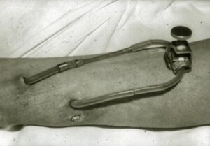
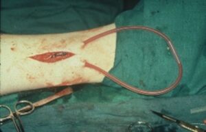
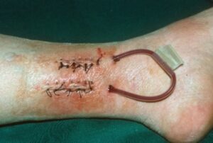
Figure: AV Shunt (a) an early AV shunt constructed in Seattle – not the metal clamp joining the arterial and venous limbs. (b) an AVshunt newly constructed before skin closure. (c) an established AV shunt. In (b) and (c) an internal stiff plastic tube joins the two limbs, with tape for added ‘security’.
Alas using the AV shunt as long term vascular access was not as easy as that brief description might imply. The shunt was prone to spontaneous thrombosis, patients were taught to watch the shunt carefully, and when loss of flow was seen, the shunt had to be de-clotted as soon as possible. Thrombosis became more common with time as venous stenosis developed. The declotting procedure was miserable, tedious and often very painful. Fine flexible sterile catheters filled with heparinised saline were inserted in the arterial and venous limbs and thrombus was aspirated. The longer the shunt had been thrombosed the more adherent was the thrombus, and the more painful and less successful was the procedure. Attempts to de-clot AV shunts with this technique were the bane of life for junior medical staff on renal units in the 1960s and early 1970s. And also for home HD patients who with their carer must themselves attempt the declotting procedure. Declotting was only endurable because of the alternative grim prospect that dialysis might no longer be possible if it failed.
Infection risk with an AV shunt was considerable given the long term placement of plastic breaching skin. With time venous stenosis became severe enough that it was necessary to re-site the venous limb more proximally. Sooner or later maintaining the shunt became impossible and a fresh shunt was placed at another ankle or forearm site. Someone receiving maintenance HD for a number of years might well run out of sites for shunt placement.
AV Fistula
The next transforming and long lasting development was the invention of the arteriovenous (AV) fistula, reported by two New York physicians, James Cimino & Michael Brescia with the vascular surgeon, Kenneth Appel. (New Engl J Med. 1966; 275:1089-92).
Typically the first choice for the AV fistula was anastomosis (side-to-side or end-to-side) of the cephalic vein to the radial artery. The vein wall proximal to the anastomosis gradually arterialised being subjected to arterial pressure, and that thickened wall became suitable for recurrent needling. Once ‘matured’ the benefits of a successful AV fistula were many: using wide bore needles higher flows could be achieved than with an AV shunt, infection risks were lower, and a good fistula might provide vascular access for years. Placement of wide bore needles by staff or patient was surprisingly well tolerated, often without local anaesthetic.
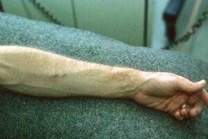
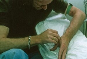
Figure: (a) An established radiocephalic AV fistula (b) patient needling his fistula
A single needle system had the attraction of less needle placement although was often allowed less efficient dialysis.
The disadvantages of the AV fistula were also well recognised. It took a few weeks to ‘mature’ so unlike the AV shunt could not be used immediately; not a significant issue when there could be a planned dialysis start, and there were sufficient available surgical resources to ensure timely AV fistula construction. For the urgent HD start, a venous catheter was necessary for vascular access, but in principle for the shortest time possible until an AV fistula was established and ready to use. A further disadvantage, significant for more women than men, is the cosmetic appearance as progressively with time firm dilated arterialised veins become more visible across the forearm. An infection risk remained with regular needling, but with good infection prevention technique this proved uncommon. In the longer term venous hypertension and more commonly venous stenosis would compromise efficiency and need surgical or radiological intervention.

Figure: Venous hypertension complicating an AV fistula
Although not perfect, the obvious superiority of the AV fistula to the AV shunt meant that by the mid-1970s the fistula had almost completely replaced the AV shunt as the first line vascular access technique.
AV graft
Another alternative to the AVF was the AV graft, which was much more widely used in the USA than the UK and most other countries. A length of PTFE woven graft was anastomosed between a suitable artery and vein. The advantage was that the graft itself could almost immediately be needled, minimising the use of venous catheters at the start of dialysis. In the USA in the 1980s and 1990s the AV graft assumed great popularity as primary vascular access. Happily this was not replicated in the UK, since the downsides of the AV graft were soon apparent, including a higher risk of infection than the AV fistula, and of aneurysmal dilatation and stenosis requiring further surgical revisions. These disadvantages were increasingly apparent in the USA by the turn of the century, and a vigorous national campaign for ‘Fistula First’ was necessary to improve practice. (The AV graft has a role, but only in complex vascular access procedures where more straightforward vascular anastomoses have been unsuccessful).
A perfect storm – vascular access strategy under pressure in the UK
In the UK although notionally our priority was ‘Fistula First’, in practice a variety of circumstances conspired to produce a ‘perfect storm’ of second best vascular access, both for individual patients, and for whole units. In the UK this was at its worst in the last two decades of the twentieth century.
Factors at play included:
- A rapidly growing dialysis population
- Continuing high rates of ‘crash landers’, those who required urgent dialysis on their first clinical contact with nephrology services
- Limited availability of vascular access surgical expertise
- A changing demographic increasing the difficulty in constructing successfully a radiocephalic fistula
- Routine use of subclavian venous catheterisation until the risks of subsequent stenosis drove a change to the routine catheterisation of internal jugular vein.
- A high risk of bacteraemia with venous catheter use, rising further with prolonged catheter placement.
- Limited availability of interventional radiological expertise for AV fistula salvage.
- Reluctance in some centres to consider peritoneal dialysis (PD) as a modality for an urgent dialysis start.
The higher morbidity and mortality of ‘crash landers’ was well recognised and multifactorial but for an important group of patients starting HD, often older and frailer, recurrent venous catheter infection requiring multiple admissions prevented successful rehabilitation with continuing HD, and contributed to a downward spiral of increasing frailty and earlier death.
The net effect was a morale-sapping negative influence on HD practice in the UK. Too many patients started HD urgently using a venous catheter which remained in use for weeks or months on end. Apart from the less efficient dialysis achieved compared to that using a well-matured AV fistula, bacteraemias were very common, and much in-patient bed capacity on renal units was consumed by patients with catheter-related problems.
The introduction of the antibiotic (usually gentamicin) ‘lock’ into catheter care was first reported in a clinical trial by Richard Fluck and colleagues in Derby (Kidney Int. 2004; 66:801-5). The results were impressive, infection rates dropping by three quarters. From the time their findings were first presented at a Renal Association meeting in 2003, its use and benefits became widespread across the UK.
‘Difficult’ vascular access
Improved management of catheter-related infection did not obviate the urgent necessity to establish permanent access using an AV fistula in as many patients as possible. But the change in the demographics and co-morbidity of the dialysis made the task for vascular access surgeons more challenging. The ‘typical’ dialysis patient of earlier days was a young or middle aged patient with large veins and good arteries. With good surgical technique high primary patency rates for AV fistulas and good longevity were achieved. But gradually the dialysis population became older with more comorbidity. Arterial calcification often made distal arteries unusable, multiple venous occlusions, related to IV cannulations during previous illnesses, would further limit the options. ‘Straightforward’ primary radiocephalic fistula formation, suitable work for trainee surgeons, became less common. Brachiocephalic fistulas and other more esoteric techniques were increasingly required, demanding more consultant surgical time. More patients required multiple access procedures. In a significant minority it was impossible to construct a usable AV fistula.
In the early days of dialysis, nephrologists were adept at placing AV shunts and a minority also learned to establish AV fistulas, for example Des Oliver in Oxford, with the obvious advantage that delays in obtaining surgeon and theatre time could be obviated. But the volume of access work, and the decreasing suitability of radiocephalic AV fistulas as primary access, inevitably squeezed out the contribution of nephrologists, and instead a collaborative partnership with vascular surgeons as well as vascular radiologists was necessary for optimal vascular access care.
National Vascular Access Survey 2006
In 2006, Kidney Research UK (KRUK) and the Renal Association (RA) combined to run a Vascular Access Survey across UK dialysis units. Kidney Research UK mainly sought information from the survey to establish research priorities, the Renal Association expected that comparative information gathered could provide support to local renal units pushing for additional vascular access resource. Unfortunately the use of information from the survey went somewhat awry. It was agreed before the survey was conducted that any emerging evidence of substandard vascular access practice would be shared first with the relevant renal unit directors for verification and for use within local clinical governance frameworks as a vehicle for change. Unfortunately a member of the senior staff at KRUK took it upon himself to write directly to the MPs in whose constituencies poor vascular access practice had been identified, but not yet verified. There was understandable ire among the clinical directors whose services had been ‘outed’ as of poor quality without data verification, and among the RA leadership who had given specific assurances to clinical directors about how the survey data would be managed. This was a rare mis-step in the working relationship between KRUK and RA, which before and since has been harmonious. KRUK moved promptly to discipline and then dismiss the director concerned, and good relations were soon restored.
Auditing vascular access
The UK Renal Registry has been auditing vascular access for the last fifteen years. It collects data on vascular access used at first dialysis in incident patients, and vascular access being used in prevalent patients. Ambitious attempts to audit the vascular access ‘pathway’ in more detail to help drive improvements in care – gathering for example the date of referral to a vascular access surgeon, the date of surgical procedures and radiological interventions – have proved impractical.
Factors improving vascular access
From the nadir of twenty years ago the vascular access challenge has came under better control. Several factors are at play:
- The number of crash landers has gradually reduced, not least due to increased awareness and improved early detection of chronic kidney disease (CKD) in primary and secondary care, ensuring a larger proportion of those who will need dialysis do reach a nephrologist in time to allow pre-emptive vascular access placement.
- Vascular access planning is prioritised. Many units identify a ‘vascular access coordinator’ (often a senior nurse) who oversees the logistics of coordination between nephrologist, dialysis unit staff, vascular surgeon and vascular radiologist, minimising the missing records, missed appointments and other apparent trivia which slow the patient’s progress through the system.
- Vascular access surgery capacity has gradually increased.
- There is more and appropriate early use of PD.
Where are we in 2026?
However over the last decade despite clinical focus and service prioritisation, the use of AV fistula has plateaued – for example 37% in incident patients, 60% in prevalent patients in the 2022 Registry Report. The problem of vascular access will not disappear, and will remain a major challenge for some patients. There will continue to be a small minority of long term HD patients who run out of access. But things are certainly better than they were.
John Feehally
Last Updated on May 30, 2026 by John Feehally